Park X-Ray
Enhance The Way You View Your Patients
i-CAT Utilization Cases
i-CAT FOR IMPLANT SURGERY
-
Select the exact implant required
-
Achieve distortion free, unmagnified measurements of the nerve canal position
-
Measure buccal/lingual width
-
Complete 3-D information to optimize treatment planning and placement for implant
-
Locate critical anatomy and determine if bone grafting or sinus lift is warranted
-
View impacted molars, cuspids, adjacent roots and other anomalies
-
Can be utilized with implant planning software
-
iCAT takes the imaging of the Temporomandibular Joint to a new level
-
The 3D image is the clearest seen to date of TMJ anatomy
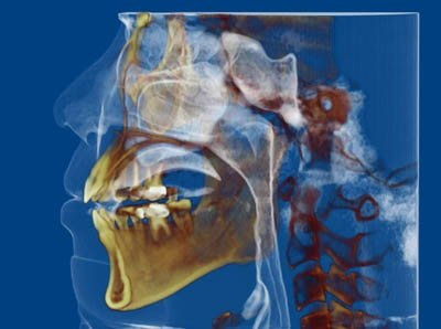
i-CAT FOR THE ORTHODONTIST
-
Orthodontists are able to create a complete orthodontic work-up from one scan including Cephalometrics, SMV, Airway, Panoramic, TM Joints, Impactions, and Facial Soft Tissue
-
See the location of impactions
-
Only a 3D scan can demonstrate the exact individual anatomy, define anatomical structures and motivate the discussions that lead to the patients' understanding of their unique clinical circumstances
i-CAT FOR MAXILLOFACIAL AND ORAL SURGEONS
-
View teeth, airway, temporomandibular condyles, the mandibular complex and sinuses
-
Visualize impactions within the alveolar bone, location relative to adjacent teeth, roots, and proximity to vital structures
-
Accurate information resulting in less invasive surgery
-
Diagnosis of bone morphology, joint space and function for TMJ analysis
-
Thorough 3-D information to optimize treatment planning